fatty liver

Fatty Liver Disease in Ahmedabad: Causes, Warning Signs & When You Must See a Gastroenterologist

Introduction

Fatty liver disease has quietly become one of the most prevalent health conditions in Ahmedabad — and most people who have it do not know. It shows no symptoms in its early stages. It does not cause pain. It is not detected by a blood test alone. It is discovered, more often than not, as an incidental finding on an ultrasound done for an entirely different reason — and by that time, it may already have been present and progressing for years. This is the silent epidemic that is affecting an estimated 1 in 3 urban Indian adults in 2026. Driven by sedentary lifestyles, calorie-dense diets, rising rates of diabetes and obesity, and increasing metabolic syndrome in Ahmedabad's working population — Non-Alcoholic Fatty Liver Disease (NAFLD) is now the leading cause of chronic liver disease in India. This guide covers what fatty liver disease is, the stages of progression, the warning signs that should never be ignored, and how Dr. Hamik Chandra Patel at Shaleen Hospital, Sola, Ahmedabad diagnoses and treats it. fatty liver

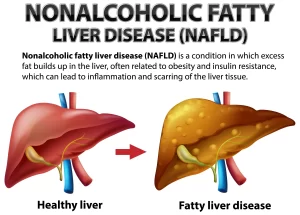
What Is Fatty Liver Disease?

Fatty liver disease occurs when excess fat accumulates in the liver cells. The liver normally contains a small amount of fat — but when fat comprises more than 5–10% of the liver's total weight, it is classified as a fatty liver. There are two main types:

NAFLD — Non-Alcoholic Fatty Liver Disease

Fat accumulation in the liver in people who drink little or no alcohol. NAFLD is the most common liver condition in India, directly linked to metabolic factors: obesity, diabetes, high cholesterol, high triglycerides, and insulin resistance.

AFLD — Alcoholic Fatty Liver Disease

Fat accumulation caused by regular heavy alcohol consumption. The liver's capacity to metabolise alcohol is exceeded, and fat accumulates. In Ahmedabad, NAFLD is by far the more prevalent — affecting middle-class, non-drinking, vegetarian adults who consume a diet high in refined carbohydrates, sugar, and fried foods.

The 4 Stages of NAFLD — Understanding Progression

NAFLD is not a single condition — it is a spectrum that progresses through four stages if not addressed:

Stage 1: Simple Fatty Liver (Steatosis)

Fat accumulates in liver cells but there is no inflammation or damage. Completely reversible with lifestyle changes. No symptoms. Often found incidentally on ultrasound.

Stage 2: NASH (Non-Alcoholic Steatohepatitis)

Fat accumulation with active inflammation and early liver cell damage. This is the dangerous transition point — NASH can progress to fibrosis and cirrhosis. May still be asymptomatic or present with mild fatigue.

Stage 3: Fibrosis

Repeated inflammation causes scar tissue (fibrosis) to form in the liver. Liver function starts to decline. Some reversibility remains with aggressive treatment.

Stage 4: Cirrhosis

Extensive scarring replaces normal liver tissue. Liver function is significantly compromised. Largely irreversible — risk of liver failure and liver cancer. Symptoms become apparent and serious. The critical insight: Most patients are diagnosed at Stage 1 or 2 — when the condition is completely manageable and reversible. This is exactly why recognising risk factors and getting evaluated is so important.

Who Is at Risk? Fatty Liver Risk Factors in Ahmedabad

Risk Factor How It Contributes
Obesity (BMI > 25–30) Most powerful risk factor — excess fat overloads liver metabolism
Type 2 Diabetes Insulin resistance drives fat deposition in liver cells
High triglycerides Elevated blood fats directly increase liver fat storage
High LDL cholesterol Metabolic dysregulation associated with NAFLD
Metabolic syndrome The combination of obesity, diabetes, hypertension, and dyslipidaemia
Sedentary lifestyle Reduces fat metabolism and increases insulin resistance
High-sugar, high-carb diet Refined flour, sugar, and sweetened beverages are converted to liver fat
Rapid weight loss Paradoxically, crash dieting or bariatric complications can worsen NAFLD
PCOD / hormonal disorders Women with PCOD have elevated NAFLD risk
Hypothyroidism Thyroid dysfunction associated with metabolic NAFLD
Age 40–60 Risk increases with age as metabolic rate declines

Warning Signs of Fatty Liver Disease — When Your Liver Is Struggling

The frustrating reality of NAFLD is that early stages produce no symptoms. But as the condition progresses — particularly to NASH, fibrosis, or cirrhosis — symptoms begin to emerge:

Early-to-Intermediate Symptoms (Stage 2–3)

  • Fatigue — persistent, unexplained tiredness not explained by other causes
  • Upper right abdominal discomfort — a dull ache or heaviness under the right rib cage (where the liver sits)
  • Mild nausea — particularly after fatty meals
  • Reduced appetite — feeling full quickly

Advanced Symptoms (Stage 3–4: Fibrosis/Cirrhosis)

  • Jaundice — yellowing of skin and whites of eyes (serious sign)
  • Abdominal swelling (ascites) — fluid accumulation in the abdomen
  • Leg swelling (oedema)
  • Easy bruising or bleeding — liver's clotting function failing
  • Itching — bile salt accumulation in the skin
  • Confusion or difficulty concentrating (hepatic encephalopathy) — toxins not cleared by the compromised liver
  • Dark urine and pale stools
If you have any of the advanced symptoms listed above, seek medical care immediately.

Why May 2026 Is World Digestive Health Month — Act Now

May is globally recognised as World Digestive Health Month — an annual initiative by the World Gastroenterology Organisation to raise awareness about preventable digestive diseases. NAFLD is one of the headline conditions for 2026 — reflecting the global recognition of its epidemic scale. For Ahmedabad residents who have been told their ultrasound shows a "mildly echogenic liver" or "fatty changes" — or who have risk factors but have never been evaluated — May is the ideal time to book a specialist consultation.

Diagnosis of Fatty Liver Disease at Dr. Gastro's Clinic, Ahmedabad

1. Blood Tests

  • Liver function tests (LFTs): ALT and AST elevation suggests liver inflammation (NASH)
  • Lipid profile: Triglycerides and cholesterol assessment
  • Fasting glucose and HbA1c: Diabetes and insulin resistance screening
  • CBC and coagulation studies: Assessing liver function in advanced disease
  • Note: Normal LFTs do not rule out NAFLD — many patients with fatty liver have completely normal blood tests

2. Ultrasound Abdomen

First-line, non-invasive test. Identifies fat accumulation as "increased echogenicity" of the liver. Can also detect advanced fibrosis signs and complications like ascites. Cannot reliably distinguish steatosis from NASH.

3. FibroScan (Transient Elastography)

A non-invasive, painless test that measures liver stiffness — indicating the degree of fibrosis. Far more informative than ultrasound for staging NAFLD. Dr. Hamik uses FibroScan at Shaleen Hospital to stage liver disease without the need for biopsy in most cases.

4. Liver Biopsy

The gold standard for definitive NAFLD staging. A small tissue sample is taken from the liver under ultrasound guidance and examined under microscope. Reserved for cases where non-invasive tests are inconclusive or where staging will significantly alter management.

5. MRI / CT Scan

Used in selected cases for detailed liver assessment and to rule out complications.

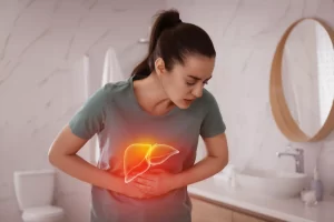
Treatment of Fatty Liver Disease at Dr. Gastro's Clinic

The Single Most Important Treatment: Weight Loss

For overweight patients with NAFLD, a 5–10% reduction in body weight consistently reduces liver fat, decreases inflammation (NASH), and can reverse early fibrosis. This is the most evidence-based intervention available for NAFLD. Dr. Hamik works with patients on a structured, medically supervised weight management programme — combining dietary modification, physical activity, and metabolic optimisation.

Dietary Modifications Specific to Ahmedabad Patients

  • Eliminate or drastically reduce: refined flour (maida), white rice, sugar, sweetened beverages, fried snacks (farsan, chakli, sev), and ultra-processed foods
  • Increase: vegetables, fruits, whole grains, legumes, nuts, and healthy fats (olive oil, nuts)
  • Follow a Mediterranean-style diet — the most evidence-backed dietary pattern for NAFLD reduction
  • Avoid: fruit juices, packaged foods labelled "low fat" (often high sugar), and alcohol

Exercise

Regular physical activity — both aerobic exercise (150 minutes/week) and resistance training — reduces liver fat independently of weight loss. Even modest increases in daily activity significantly improve NAFLD.

Managing Associated Conditions

  • Diabetes control: HbA1c optimisation reduces liver fat
  • Lipid management: Statins, fibrates as appropriate
  • Blood pressure control
  • Thyroid optimisation

Medications

While there is currently no FDA-approved medication specifically for NAFLD, Dr. Hamik uses evidence-based pharmacological approaches including:
  • Vitamin E — for non-diabetic NASH patients
  • Pioglitazone — for NASH with diabetes/prediabetes
  • GLP-1 analogues (semaglutide, liraglutide) — showing strong benefit in NAFLD/NASH with obesity
  • New NASH-specific agents currently in Phase 3 trials

Regular Monitoring

Even patients who respond well to lifestyle modification require regular monitoring — blood tests every 6 months, annual ultrasound or FibroScan — to ensure disease regression and detect any recurrence.

Conclusion

Fatty liver disease is not a condition to accept as "normal" or dismiss as a minor finding on your ultrasound report. It is a progressive liver disease that, without intervention, can advance to cirrhosis and liver failure — conditions that have no cure. The good news is that at Stage 1 and Stage 2, fatty liver is one of the most reversible conditions in medicine. With the right guidance, appropriate weight management, and targeted treatment of metabolic risk factors, most patients achieve significant liver improvement within 12–24 months. At Dr. Gastro's clinic at Shaleen Multi Speciality Hospital, Sola, Ahmedabad, Dr. Hamik Chandra Patel provides comprehensive fatty liver treatment in Ahmedabad — from FibroScan-based staging to personalised, evidence-based treatment plans. 📍 Science City Road, Sola, Ahmedabad – 380060 📞 9512039041 / 8238092233 Book Your Liver Assessment → drgastro.in/contact-us/
Read more...

Appendix Pain or Gas Pain? How to Identify Appendicitis Symptoms & Why Early Surgery Matters

Introduction

One of the most common medical emergencies in Ahmedabad is also one of the most frequently misdiagnosed: appendicitis. Every week, patients arrive at Shaleen Hospital in Sola who have been managing what they assumed was gas pain or indigestion for 12–24 hours — only to discover that their appendix is acutely inflamed and at risk of rupturing. This delay is dangerous. A ruptured appendix can cause life-threatening peritonitis and sepsis. The challenge is that early appendicitis genuinely feels similar to gas pain, period cramps, or ordinary stomach upset. This guide helps you tell the difference — and explains exactly when abdominal pain requires immediate medical attention.

What Is the Appendix?

The appendix is a small, finger-shaped pouch attached to the large intestine in the lower-right abdomen. In adults, it measures approximately 8–10 cm. Its exact function is debated — it appears to have some role in immune function in early childhood but is not essential in adults. Appendicitis occurs when the appendix becomes inflamed — usually because its opening becomes blocked by hardened stool, mucus, or in some cases, an infection. Bacterial growth inside the blocked appendix causes swelling, increased pressure, and if untreated — perforation (rupture). Drgastro

Gas Pain vs Appendicitis: How to Tell the Difference

This is the critical question most patients ask — and the answer lies in understanding the very specific characteristics of appendicitis pain.

Gas Pain — Characteristics

  • Location: Anywhere in the abdomen, often crampy and moving
  • Character: Comes and goes in waves; relieved by passing gas or having a bowel movement
  • Associated with: Bloating, burping, gurgling sounds, bowel urgency
  • Duration: Minutes to a few hours — then resolves
  • Aggravating factors: Eating certain foods, irregular meals, constipation
  • Relief: Passing gas, antispasmodic medication, bowel movement
  • Fever: No fever — gas pain does not cause fever

Appendicitis Pain — Characteristics

  • Location: Starts around the navel (umbilicus) and migrates to the lower-right abdomen (McBurney's point) within hours
  • Character: Constant, worsening — not relieved by gas or bowel movement
  • Onset: Gradual onset — mild at first, progressively intensifying over hours
  • Duration: Does not resolve — worsens over 12–24 hours without treatment
  • Movement: Pain worsens with movement, coughing, or pressing and releasing the abdomen
  • Associated with: Fever (38–39°C), nausea, vomiting, loss of appetite
  • Key sign: Rebound tenderness — pain that is worse when pressure on the lower-right abdomen is suddenly released
  • Relief: Nothing relieves it — antacids, antispasmodics, and passing gas do NOT help

The Classic Progression of Appendicitis

Understanding how appendicitis typically develops helps you recognise when to seek urgent care: Hours 1–6: Vague Abdominal Discomfort Diffuse, dull pain around the navel. Many patients mistake this for indigestion, gas, or overeating. Nausea and loss of appetite are common early signs. Hours 6–12: Pain Migrates to Lower Right Abdomen This is the hallmark of appendicitis — pain shifting to McBurney's point (one-third of the way from the right hip bone to the navel). The pain becomes more constant, sharper, and localised. Hours 12–24: Intensifying Pain, Fever Pain is now severe and constant. Low-grade fever (38–39°C) develops as infection worsens. Movement, coughing, and pressing the abdomen become acutely painful. Beyond 24–72 Hours: Risk of Rupture Without treatment, the inflamed appendix can rupture — spilling infected contents into the abdominal cavity. This causes peritonitis — diffuse, severe abdominal pain, high fever, and sepsis. Ruptured appendicitis requires more complex surgery and longer recovery.

Warning Signs That Require IMMEDIATE Medical Attention

Go to the emergency department at Shaleen Hospital immediately if you have:
  • Severe pain in the lower-right abdomen that has persisted for more than 4–6 hours
  • Pain that started around the navel and has shifted to the lower right
  • Abdominal pain accompanied by fever (above 38°C)
  • Nausea and vomiting with lower-right abdominal pain
  • Pain that WORSENS when you release pressure after pressing your lower-right abdomen
  • Pain so severe you cannot stand upright or walk normally
  • Sudden relief of severe pain followed by worsening diffuse pain (sign of rupture)
Do NOT eat, drink, or take painkillers before being evaluated by a doctor — these can mask symptoms and delay diagnosis.

Diagnosis of Appendicitis at Dr. Gastro's Clinic

Dr. Hamik Chandra Patel uses a combination of clinical assessment and investigations to diagnose appendicitis:

Clinical Examination

  • McBurney's Point tenderness — direct tenderness at the classic appendicitis location
  • Rebound tenderness (Blumberg's sign) — pain worse on pressure release
  • Rovsing's sign — pain in the right when the left abdomen is pressed
  • Psoas sign — pain when the right hip is extended (posterior appendix)
  • Obturator sign — pain with hip rotation (pelvic appendix)

Blood Tests

  • Complete Blood Count (CBC): Elevated white blood cell count (leucocytosis) indicates infection
  • CRP (C-Reactive Protein): Elevated in inflammation

Ultrasound Abdomen

First-line imaging in Ahmedabad. Can visualise an inflamed appendix in many cases, and rules out other causes (ovarian cysts, kidney stones, etc.)

CT Scan Abdomen

Gold standard when diagnosis is uncertain. Provides the most accurate imaging of the appendix and detects perforation or abscess formation.

Alvarado Score

A clinical scoring system using symptoms, signs, and blood tests to calculate appendicitis probability — guiding the decision to operate.

Treatment: Laparoscopic Appendicectomy by Dr. Hamik, Ahmedabad

Appendicectomy — surgical removal of the inflamed appendix — is the definitive and only reliable treatment for acute appendicitis. There is no effective long-term medical management for an inflamed appendix.

Why Laparoscopic Appendicectomy?

Dr. Hamik Chandra Patel performs laparoscopic (keyhole) appendicectomy — the gold standard surgical approach:
  • 3 small incisions (5–10 mm each) instead of a large open cut
  • Camera and instruments inserted through the incisions
  • Appendix identified, its blood supply clipped, and it is cleanly removed
  • Procedure takes 30–45 minutes under general anaesthesia
  • Same-day or next-day discharge for uncomplicated appendicitis
  • Return to desk work within 5–7 days
  • Full recovery within 2–3 weeks
  • Minimal scarring — barely visible marks remain

Uncomplicated vs Perforated Appendicitis

Uncomplicated appendicitis (no rupture): Straightforward laparoscopic surgery with excellent outcomes. Short hospital stay, quick recovery. Perforated appendicitis (ruptured): Requires more complex surgery to clean the abdominal cavity, possible drain placement, and a longer hospital stay. Recovery is longer and risk of complications is higher. This is why timing matters enormously — the sooner the surgery, the better the outcome. gas pain

Why Early Surgery Saves Lives — and Prevents Complications

Every hour of delay between onset of appendicitis symptoms and surgery increases the risk of rupture. Studies show:
  • Appendix rupture rate increases significantly after 36 hours of symptoms
  • Ruptured appendicitis has a complication rate 3–5 times higher than uncomplicated cases
  • Ruptured appendicitis requires a 3–5 times longer hospital stay
  • Risk of post-operative complications (wound infection, abscess, prolonged ileus) is dramatically higher after perforation
The message is unambiguous: if appendicitis is suspected, get to the hospital immediately — do not wait and see if it improves.

Conclusion

Appendicitis is a time-critical surgical emergency. The difference between a simple 45-minute operation and a complex, potentially life-threatening one is measured in hours — not days. If you or a family member in Ahmedabad has lower-right abdominal pain that is constant, worsening, and accompanied by fever and nausea — do not try to manage it at home. Seek emergency evaluation immediately. Dr. Hamik Chandra Patel at Shaleen Multi Speciality Hospital, Sola is available for emergency surgical consultation and is experienced in laparoscopic appendicectomy — delivering fast, safe, minimally invasive appendicitis surgery in Ahmedabad. 📍 Science City Road, Sola, Ahmedabad – 380060 | 📞 9512039041 / 8238092233
Read more...

Acidity vs GERD: How to Tell the Difference & When to See a Gastro Surgeon in Ahmedabad

Introduction

Every second person in Ahmedabad has experienced acidity at some point. The burning sensation after a heavy meal, the sour taste in the mouth after a late dinner, the discomfort when you lie down — these are familiar experiences for millions of Indians. But here is the problem: what most people dismiss as "acidity" is often something more significant — Gastroesophageal Reflux Disease (GERD) — a chronic, progressive condition that, when left untreated, can lead to serious complications including oesophageal ulcers, Barrett's Oesophagus, and even oesophageal cancer. This guide explains the critical difference between ordinary acidity and GERD, the warning signs that mean you need a specialist rather than another antacid, and how Dr. Hamik Chandra Patel at Shaleen Hospital, Sola, Ahmedabad diagnoses and treats both conditions.

What Is Acidity (Acid Indigestion)?

Acidity — also called acid indigestion or heartburn — refers to a temporary increase in stomach acid production that causes a burning sensation in the upper abdomen or chest. It is extremely common and is usually triggered by:
  • Eating spicy, fried, or oily foods
  • Eating large meals quickly
  • Lying down immediately after eating
  • Drinking too much tea, coffee, or alcohol
  • Eating late at night
  • Stress and anxiety
Key characteristics of simple acidity:
  • Occurs occasionally — after specific triggers
  • Resolves within 1–2 hours, often with an antacid
  • Does not significantly disrupt daily life
  • No persistent symptoms between episodes
Simple acidity is a symptom, not a disease. It is managed with dietary changes and over-the-counter antacids and does not require specialist care.

What Is GERD (Gastroesophageal Reflux Disease)?

GERD is a chronic condition in which stomach acid repeatedly flows back (refluxes) into the oesophagus — the tube connecting your mouth to your stomach. Unlike simple acidity, GERD is not occasional. It is persistent, progressive, and causes measurable damage to the oesophageal lining over time. In GERD, the Lower Oesophageal Sphincter (LES) — the muscular valve that prevents acid from flowing back up — is weakened or dysfunctional. As a result, acid repeatedly escapes from the stomach into the oesophagus, irritating and damaging its lining with every episode. GERD is diagnosed when:
  • Reflux symptoms occur 2 or more times per week
  • Symptoms significantly affect quality of life
  • Or endoscopy shows evidence of acid-related damage to the oesophagus

Acidity vs GERD: The Key Differences

Feature Simple Acidity GERD
Frequency Occasional (after triggers) 2+ times per week, often daily
Duration Resolves in 1–2 hours Persistent, chronic
Response to antacids Usually resolves Partial relief or no relief
Night-time symptoms Rare Common — often wakes patients
Regurgitation Uncommon Very common (sour/bitter fluid in mouth)
Chest pain Mild, brief Can be severe and prolonged
Impact on life Minimal Significant — disrupts sleep, eating, activity
Oesophageal damage None Progressive damage over time
Requires specialist No Yes — if untreated, risks serious complications

Symptoms of GERD — What to Watch For

GERD presents with a range of symptoms, both typical and atypical:

Typical GERD Symptoms

  • Heartburn — burning sensation in the chest, occurring frequently (2+ times/week)
  • Acid regurgitation — sour or bitter taste as acid rises to the mouth or throat
  • Chest pain — especially when lying down or bending forward
  • Difficulty swallowing (dysphagia) — as the oesophagus becomes inflamed or narrows
  • Sensation of a lump in the throat (globus)

Atypical GERD Symptoms (Often Misdiagnosed)

  • Chronic cough — particularly at night, without obvious respiratory cause
  • Hoarseness or voice changes — acid irritating the vocal cords
  • Chronic throat clearing or sensation of something stuck in the throat
  • Worsening of asthma — acid aspiration triggering bronchospasm
  • Dental erosion — acid damaging tooth enamel
  • Disrupted sleep — frequent waking due to acid discomfort

Why GERD in Ahmedabad Is More Common Than You Think

Several factors specific to Ahmedabad and Gujarat's lifestyle patterns make GERD prevalence particularly high: Dietary patterns: Ahmedabad's cuisine is rich in spicy, oily, and fried foods — all major GERD triggers. Farsan, dhokla with chutney, puri shaak, and other beloved local foods are consumed in large quantities at irregular meal times. Late eating habits: Many Ahmedabad families eat dinner late — often after 9 PM — and retire to sleep within 1–2 hours. This allows reflux to occur with the added disadvantage of horizontal position. High tea and coffee consumption: Gujarat's culture of multiple cups of tea throughout the day is a consistent GERD aggravator for susceptible individuals. Sedentary lifestyle: Long working hours at desks, minimal physical activity, and stress from business pressures contribute to both oesophageal motility disorders and increased acid production. Rising obesity rates: Excess abdominal fat increases intra-abdominal pressure, directly worsening acid reflux. Ahmedabad's urban population is experiencing rising obesity rates that correlate directly with GERD incidence.

Complications of Untreated GERD

This is where GERD transitions from an inconvenience to a medical concern. Untreated or inadequately managed GERD can cause:

1. Oesophagitis

Chronic acid exposure causes inflammation and ulceration of the oesophageal lining. Symptoms intensify and healing becomes more difficult without medical intervention.

2. Oesophageal Stricture

Repeated inflammation leads to scar tissue formation, narrowing the oesophagus. Swallowing becomes progressively more difficult — initially with solids, then with liquids.

3. Barrett's Oesophagus

One of the most serious GERD complications. Repeated acid damage causes the normal oesophageal lining to be replaced by abnormal intestinal-type cells — a precancerous condition. Patients with Barrett's require regular endoscopic surveillance.

4. Oesophageal Cancer

Long-standing Barrett's Oesophagus can progress to oesophageal adenocarcinoma — one of the fastest-rising cancers globally. Early detection through regular endoscopy is the only reliable protection.

5. Aspiration Pneumonia

In severe GERD, acid and food particles can be aspirated into the lungs during sleep, causing pneumonia.

When to See Dr. Gastro in Ahmedabad for GERD

Stop relying on antacids and see Dr. Hamik if:
  • Your heartburn or reflux occurs 2 or more times per week
  • Symptoms persist despite taking over-the-counter antacids regularly
  • You wake up at night with acid coming up
  • You have difficulty swallowing — even occasionally
  • You are losing weight without trying
  • You have a persistent cough that has not responded to respiratory treatment
  • You have had acidity symptoms for more than 3–5 years without proper evaluation
  • You are over 45 and have new-onset reflux symptoms
  • There is any blood in your vomit or stools
These are not signs that you need more antacids. These are signs that you need a gastroenterologist.

How Dr. Hamik Diagnoses GERD in Ahmedabad

1. Clinical Evaluation

A detailed history of symptom frequency, triggers, diet, and lifestyle — plus examination for associated conditions (hiatal hernia, obesity).

2. Upper Endoscopy (Gold Standard)

Endoscopy provides direct visualisation of the oesophagus, stomach, and duodenum. It identifies oesophagitis severity, Barrett's Oesophagus, strictures, ulcers, and hiatal hernia. Biopsies are taken if Barrett's is suspected.

3. 24-Hour pH Monitoring

A thin probe placed in the oesophagus measures acid levels over 24 hours — the most precise way to confirm and quantify GERD.

4. Oesophageal Manometry

Measures the pressure and function of the oesophagus and LES — important when surgical treatment for GERD is being considered.

5. Barium Swallow X-ray

An older technique still useful in specific cases — particularly when structural abnormalities of the oesophagus are suspected.

GERD Treatment Options at Dr. Gastro's Clinic, Ahmedabad

Lifestyle Modifications (First Line — Essential for All Patients)

  • Elevate the head of the bed by 15–20 cm
  • Eat smaller meals more frequently — avoid large meals
  • Do not lie down for 2–3 hours after eating
  • Avoid known trigger foods (spicy, fried, oily, tomatoes, citrus, chocolate, mint)
  • Reduce or eliminate tea, coffee, and alcohol
  • Lose weight if overweight — even 5–10% weight loss significantly reduces GERD
  • Stop smoking — smoking weakens the LES directly

Medical Treatment

  • Proton Pump Inhibitors (PPIs): Omeprazole, pantoprazole, rabeprazole — the most effective medications for reducing acid production. Most GERD patients respond well to a properly dosed PPI regimen.
  • H2 Receptor Blockers: Ranitidine-equivalent agents — used for milder GERD or as add-on therapy.
  • Prokinetic Agents: Improve oesophageal motility and speed gastric emptying in selected patients.
  • Antacids: For immediate symptom relief only — not for long-term GERD management.

Surgical Treatment — Anti-Reflux Surgery (Fundoplication)

For patients who do not respond adequately to medication, have severe complications, or wish to avoid lifelong medication, laparoscopic fundoplication (Nissen or partial) is the surgical solution. Dr. Hamik Chandra Patel performs laparoscopic anti-reflux surgery at Shaleen Hospital with excellent results — eliminating acid reflux by strengthening the LES with a minimally invasive technique. Laparoscopic fundoplication results in:
  • 90%+ long-term symptom control
  • Ability to discontinue PPIs in most patients
  • Short hospital stay (1–2 days)
  • Return to normal activities within 1–2 weeks

Conclusion

Acidity and GERD are not the same condition — and treating GERD as if it were simple acidity (with antacids and diet alone) is a mistake that leads to progressive oesophageal damage, increasing medication dependency, and in serious cases, precancerous changes. If you have been managing what you call "acidity" for months or years without proper evaluation — it is time to see a specialist. At Dr. Gastro's clinic at Shaleen Multi Speciality Hospital, Sola, Ahmedabad, Dr. Hamik Chandra Patel provides expert GERD treatment in Ahmedabad — from accurate endoscopic diagnosis to medical management, dietary counselling, and laparoscopic anti-reflux surgery for patients who need it. 📍 Science City Road, Sola, Ahmedabad – 380060 | 📞 9512039041 / 8238092233 Book a Consultation → drgastro.in/contact-us/
Read more...
Endoscopy vs Colonoscopy

Endoscopy vs Colonoscopy: What’s the Difference, When Do You Need It & What to Expect at Dr. Gastro, Ahmedabad

Introduction

Two of the most frequently asked questions at Dr Gastro's clinic in Ahmedabad are: "Doctor, do I need an endoscopy or a colonoscopy?" and "What exactly happens during these procedures?" Both are diagnostic tools used by gastroenterologists to examine the digestive tract from the inside. Both involve a flexible camera. But they look at entirely different parts of your digestive system — and they are recommended for very different symptoms and conditions. If your doctor has recommended an endoscopy in Ahmedabad or a colonoscopy — or if you have symptoms that might require one — this guide will tell you everything you need to know: the differences, when each is recommended, what the procedure involves, and what to expect at Dr Gastro's clinic at Shaleen Multi Speciality Hospital, Sola, Ahmedabad.

What Is an Endoscopy?

An endoscopy (also called upper GI endoscopy or gastroscopy) is a procedure that examines the upper digestive tract — the oesophagus (food pipe), stomach, and the first part of the small intestine (duodenum). A thin, flexible tube called an endoscope — with a high-definition camera and light at its tip — is passed gently through the mouth, down the throat, and into the stomach. The camera transmits live, magnified images to a monitor, allowing Dr Hamik Chandra Patel to examine the lining of your upper digestive tract in detail.

What Can an Upper Endoscopy Diagnose?

  • Acid reflux (GERD) — oesophageal inflammation, strictures
  • Gastric ulcers — stomach or duodenal ulcers
  • Helicobacter pylori infection — the bacterium responsible for most peptic ulcers
  • Oesophagitis — inflammation of the oesophagus
  • Barrett's Oesophagus — a precancerous change in the oesophageal lining
  • Hiatal hernia — stomach pushing through the diaphragm
  • Unexplained anaemia — bleeding from the upper GI tract
  • Coeliac disease — biopsy samples from the duodenum for diagnosis
  • Stomach cancer — early detection is the key to survival
  • Swallowing difficulty (dysphagia) — identifying structural causes

What Can an Endoscopy Treat?

Beyond diagnosis, an endoscopy is also therapeutic:
  • Biopsy collection for laboratory analysis
  • Polyp removal from the stomach
  • Bleeding control (endoscopic haemostasis)
  • Removal of swallowed foreign bodies
  • Dilatation of narrowed areas (strictures)
  • ERCP — examination and treatment of bile duct and pancreatic conditions

What Is a Colonoscopy?

A colonoscopy examines the lower digestive tract — the entire large intestine (colon) and the terminal portion of the small intestine (ileum). A longer, flexible scope is passed through the rectum and guided through the entire colon. This procedure requires bowel preparation the day before to ensure the colon is clean and clearly visible.

What Can a Colonoscopy Diagnose?

  • Colorectal cancer — the most important reason for colonoscopy
  • Polyps — precancerous growths on the colon lining (removed during the procedure)
  • Inflammatory bowel disease (IBD) — Crohn's disease and ulcerative colitis
  • Diverticular disease — small pouches in the colon wall
  • Unexplained rectal bleeding — identifying the source
  • Chronic diarrhoea or constipation — investigating structural causes
  • Iron deficiency anaemia — unexplained blood loss from the colon
  • Post-cancer surveillance — monitoring after previous colorectal cancer treatment

What Can a Colonoscopy Treat?

  • Polypectomy — removal of colon polyps (preventing cancer before it starts)
  • Bleeding control — endoscopic treatment of bleeding points
  • Biopsy — tissue samples for IBD diagnosis and cancer detection

Endoscopy vs Colonoscopy: Key Differences

Feature Endoscopy (Upper GI) Colonoscopy (Lower GI)
Area examined Oesophagus, stomach, duodenum Entire colon, rectum, terminal ileum
Entry point Mouth Rectum
Preparation required Fasting for 6–8 hours Bowel prep (laxatives) the day before
Procedure duration 10–20 minutes 30–60 minutes
Sedation Light sedation or throat spray Deeper sedation recommended
Primary use Upper GI symptoms (acid, pain, vomiting) Lower GI symptoms (bleeding, altered bowel habit)
Cancer screening Stomach/oesophageal cancer Colorectal cancer

When Does Dr. Hamik Recommend an Endoscopy in Ahmedabad?

Dr. Hamik Chandra Patel recommends upper endoscopy when patients present with:
  • Persistent upper abdominal pain — especially pain that does not respond to antacids
  • Chronic acidity or GERD — when symptoms persist despite medication or when Barrett's Oesophagus is suspected
  • Difficulty swallowing (dysphagia) — food sticking in the throat or chest
  • Unexplained nausea or vomiting — particularly in older patients
  • Unexplained weight loss — a red flag symptom requiring investigation
  • Vomiting blood (haematemesis) — an emergency requiring immediate endoscopy
  • Black, tarry stools (melaena) — indicating upper GI bleeding
  • Anaemia — when a bleeding source in the upper GI tract is suspected
  • Positive H. pylori test — to confirm and assess associated damage
  • Family history of stomach cancer — surveillance in high-risk individuals
  • Suspected coeliac disease — biopsy from the duodenum

When Does Dr Hamik Recommend a Colonoscopy?

Colonoscopy is recommended for:
  • Rectal bleeding or blood in stools — never ignore this symptom
  • Unexplained change in bowel habits — new constipation, diarrhoea, or alternating pattern
  • Persistent lower abdominal cramps or bloating
  • Iron deficiency anaemia — when colonoscopy is needed to rule out colorectal cancer
  • Colorectal cancer screening — recommended from age 45–50, or earlier with family history
  • Previous colorectal polyps or cancer — surveillance colonoscopy
  • Suspected inflammatory bowel disease — Crohn's disease or ulcerative colitis
  • Abnormal CT scan findings in the colon requiring direct visualisation
  • Unexplained significant weight loss combined with bowel symptoms

What to Expect During an Endoscopy at Dr Gastro's Clinic, Ahmedabad

Before the Procedure

  • Fast for 6–8 hours before your endoscopy (no food or water)
  • Inform Dr. Hamik of any medications, especially blood thinners
  • Bring a companion who can accompany you home if sedation is used
  • Routine blood tests may be requested beforehand

During the Procedure

  • You will lie on your left side on a procedure bed
  • A mouthguard is placed to protect your teeth and the scope
  • A throat spray or light sedation is administered for comfort
  • The endoscope is gently passed through your mouth — you may feel mild pressure, but should not feel pain
  • The entire procedure takes 10–20 minutes
  • If biopsies are taken or polyps removed, you will not feel this

After the Procedure

  • You will rest in recovery for 30–60 minutes
  • If sedated, you cannot drive yourself home
  • Mild bloating, belching, or a sore throat for a few hours is normal
  • Biopsy results are typically available within 5–7 days
  • Dr. Hamik discusses findings immediately after the procedure

What to Expect During a Colonoscopy at Dr Gastro's Clinic, Ahmedabad

Bowel Preparation — The Day Before

The most important part of colonoscopy preparation is ensuring your colon is completely clean. Dr Hamik's team will provide:
  • A specific bowel preparation laxative to take the evening and morning before
  • Clear dietary instructions (clear liquids only the day before)
  • Timing instructions for medication use
A clean bowel is critical — inadequate preparation leads to a repeat procedure.

During the Procedure

  • Performed under sedation for your comfort
  • Duration is typically 30–60 minutes
  • You may feel mild cramping as the scope moves through the colon's curves
  • Any polyps found are removed during the same procedure
  • Biopsies are taken from any suspicious areas

After the Procedure

  • Recovery takes 1–2 hours post-sedation
  • Mild bloating or gas for a few hours is normal
  • You cannot drive on the day of the procedure
  • Results discussed immediately; biopsy reports in 5–7 days
  • A soft diet is recommended for the remainder of the day
endoscopy vs colonoscopy

Can Both Be Done on the Same Day?

Yes — in selected cases, Dr Hamik Chandra Patel at Shaleen Hospital can perform both upper endoscopy and colonoscopy in a single session. This is called bidirectional endoscopy and avoids the need for two separate preparations and appointments. This is particularly convenient for patients with symptoms suggesting both upper and lower GI pathology, or for comprehensive screening.

Endoscopy Cost in Ahmedabad — What to Expect

The cost of endoscopy or colonoscopy in Ahmedabad varies based on whether biopsies are taken, sedation is used, and the specific procedure performed. Dr Gastro's clinic at Shaleen Hospital provides transparent cost estimates before any procedure. For specific pricing, please contact: 9512039041 / 8238092233.

Why Choose Dr Gastro for Endoscopy in Ahmedabad?

Dr Hamik Chandra Patel's Specialised Expertise: Dr Hamik is a qualified gastroenterologist and laparoscopic surgeon with specialised training in diagnostic and therapeutic endoscopy. His experience includes thousands of upper and lower GI endoscopy procedures, complex ERCP, and endoscopic polypectomy. Advanced Endoscopy Suite: Shaleen Multi-Speciality Hospital operates a fully equipped, hygienic endoscopy suite with high-definition imaging systems, ensuring the most accurate diagnostic views possible. Same-Day Report Discussion: Unlike many centres where patients wait days to speak with a doctor, Dr Hamik discusses endoscopy findings immediately after the procedure — giving you clarity and peace of mind the same day. Patient Comfort and Safety: Appropriate sedation, careful monitoring, and a caring nursing team ensure the procedure is as comfortable as possible. Post-procedure recovery is supervised until you are fully ready to leave.

Conclusion

Endoscopy and colonoscopy are among the most powerful diagnostic tools in gastroenterology — providing direct, high-definition views of your digestive tract that no blood test or ultrasound can match. When symptoms suggest digestive pathology, prompt endoscopic investigation is the fastest route to an accurate diagnosis and appropriate treatment. At Dr Gastro's clinic at Shaleen Multi Speciality Hospital, Sola, Ahmedabad, Dr Hamik Chandra Patel provides expert endoscopy services — with technical skill, the right equipment, and patient-centred communication that help you understand your diagnosis and options clearly. Do not delay investigation of persistent digestive symptoms. 📍 Shaleen Multi Speciality Hospital, Science City Road, Sola, Ahmedabad – 380060 📞 9512039041 / 8238092233 🕐 Mon–Fri: 8:30 AM – 5:00 PM | Saturday: 9:30 AM – 1:00 PM Book an Appointment → drgastro.in/contact-us/
Read more...